CHONDRODYSPLASIA PUNCTATA
Kalia Vishal, Vibhuti, Aggarwal Tanuj Dyanand Medical College & Hospital, Ludhiana - 141001, India Address for Correpondence: Dr. Tanuj Aggarwal, 186-B, Kendriya Vihar, Sector 48- B, Chandigarh 160047. E-Mail: tanujagg@gmail.com
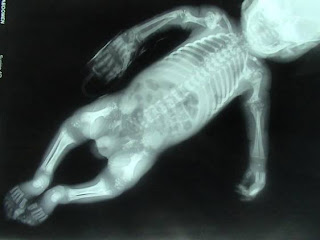
A preterm, small for gestational age infant (2 kilograms) born at 36 weeks 4 days to a gravida five mother by normal vaginal delivery presented on day five with continuous fever (102 degree F) associated with poor feeding and lethargy. The antenatal history revealed polyhydramnios on ultrasonography performed at five and seven months. The anthropometric parameters of the infant were as follows - weight: 2 kilograms, height: 42 cms, head circumference: 31 cms, arm span: 35 cms. The physical examination revealed open anterior and posterior fontanelle, cleft soft palate, short femora and humeri, deformed knee and elbow joints and overriding of skull sutures. The infantogram showed stippling of the long bone and vertebral epiphyses. (Figure 1) suggestive of chondrodysplasia punctata.
Figure 1: Infantogram showed stippling and punctate calcification of the long bone and vertebral epiphyses
Chondrodysplasia punctata is a rare, multisystem, developmental disorder characterized by the presence of stippled foci of calcification in hyaline cartilage, coronal vertebral clefting, dwarfing, joint contractures, congenital cataract, icthyosis and severe mental retardation. The chondrodysplasia punctata family includes a dominant autosomal form (Conradi-Hunnermann Disease), a recessive autosomal form (rhizomelic), forms linked to X recessive (chondrodysplasia X 1) and dominant (chondrodysplasia X 2 or Conradi Hunnermann-Happle). During the nineties two milder forms were described, tibial metacarpal and brachytelephalangic. In 1991, Boronchowitz described a patient with an apparently new syndrome of chondrodysplasia punctata, distinct from previously described forms. He suggested the term "chondrodysplasia punctata, humero-metacarpal (HM)" type. In contrast, with the other forms, the rhizomelic form has a bad prognosis, with repeated respiratory infections and death during the first two years of life. The rhizomelic form of chondrodysplasia is rare, with 72 cases described in literature by 1995. Despite the number and structure of peroxisomes being normal, a functional defect of these organelles result in an enzyme deficiency, characterized by decrease in the synthesis of plasmalogen, a reduction in the oxidation of phytanic acid and of the presence of an unprocessed peroxisomal hepatic enzyme, 3-oxoacyl-CoA-thiolase. Plasmatic phytanic acid is found to be increased and the synthesis of plasmalogen in the fibroblasts and their content in the erythrocytes are reduced. Chromosome study denotes a mutation in PEX 7 gene and more than 20 mutations have been described. A DHAPAT deficiency (acyl-CoA-dihydroxyacetone phosphate acyltransferase) results in rhizomelic chondrodysplasia punctata subtype 1. Subtypes 2 and 3 are caused by a reduction in DHAPAT activity and alkyl-DHAP synthesis, respectively. Radiologic criteria for diagnosis of in the young infant, in late infancy and in childhood was described by Gilbert EF et al (1976). These criteria in a young infant include radiolucent coronal clefts dividing all or most of the thoracic and lumbar vertebral bodies, short and wide humeri with flared metaphyses and punctate calcification at various areas including the entire spine, sacrum, hands, feet, trachea and thyroid, symmetrical brachymetacarpals especially of the 4th metacarpal and hypoplastic distal phalanges. In late infancy and childhood the radiologic criteria included demineralization in all bones with slow maturation, flat vertebral, bodies, short humeri and femora, metaphyseal flaring, especially in the distal humerus, proximal femur and tibia, immature shape of pelvis and disappearance of the punctate calcification with advancing age. Cataracts are present in about 72% of cases and skin changes in about 27%. Happle (1981) suggested that cataracts are present in about 2/3 of the rhizomelic and x-linked dominant forms. In the rhizomelic form, the opacities tend to be bilateral and symmetric in the x-linked form, they are usually asymmetric and often unilateral. Currently, the rhizomelic chondrodysplasia punctata diagnosis is made based on clinical characteristics which are compatible with the syndrome, punctate calcifications, rhizomelia, and the biochemical findings which include serum phytanic acid assay and investigation of plasmalogen synthesis in a fibroblast culture. However, there are descriptions of cases which have been diagnosed purely by radiological and clinical criteria. The clinical characteristics include ichthyosis, irregular foci of alopecia, feeble skin, shortened neck, flattened facies with saddle nose, permanently flexed knee and elbow joints, besides limbs shortening, microcephaly and micrognathia and history of deglutition difficulty. The differential diagnosis includes other causes for chondrodysplasia punctata, Keutel syndrome Zellweger syndrome, Smith-Lemli-Opitz syndrome, neonatal Refsum disease, neonatal adrenoleucodystrophy, neonatal lupus, trisomy 21 or 18, fetal alcoholic syndrome, congenital infections and maternal use of coumarin-like compounds or phenytoin during gestation.
Reference
Borochowitz Z. Generalized chondrodysplasia punctata with shortness of humeri and brachymetacarpy: humero-metacarpal (HM) type: variation or heterogenecity- Am. J. Med. Genet 1991; 41(4): 417-422.
Fourie DT. Chondrodysplasia punctata: case report and literature review of patients with heart lesions. Pediatr Cardiol 1995; 16: 247-250.
Gilbert E F, Opitz J M, Spranger J W, Langer L O Jr, Wolfson J J, Viseskul C. Chondrodysplasia punctata - rhizomelic form. Pathologic and radiologic studies of three infants. Eur J Pediatr 1976; 123(2): 89-109.
Heymans HS, Oorthuys JW, Nelek G, Wanders RJ, Schutgens RB. Rhizomelic chondrodysplasia punctata: another peroxisomal disorder [letter]. N Engl Med.1985; 13: 187-188,
Kumada S, Hayashi M, Kenmochi J, et al. Lethal form of chondrodysplasia punctata with normal plasmalogen and cholesterol biosynthesis. Am J Med Genet 001; 98: 250-255.
Pascolat G, Zindeluk J L, Abrao K C, Rodrigues F M, Guedes C I. Rhizomelic chondrodysplasia punctata. J. Pediatr (Rio J). 2003;79(2): 189-192. Latest Updated On: 1st December 2008 Vol 5 Issue 12 Art # 50Advance Access: 1st April 2008
How to cite this url
Vishal K,Vibhuti, Tanuj A . Chondrodysplasia Punctata. Pediatric Oncall [serial online] 2008 [cited 2008 December 1];5. Art # 50. Available from:
http://www.pediatriconcall.com/fordoctor/viewersChoice/chondrodysplasia_punctata.aspAdvance Access: 1st April 2008


Nenhum comentário:
Postar um comentário